Monthly Archives :

March 2023

BLOAT/ RUMINAL TYMPANY

BLOAT/ RUMINAL TYMPANY 1280 720 Kingsley Emmanuel Bentum

Bloat is an uncomfortable condition in ruminants marked by an over distension of the rumen (the first of the four divisions of stomach in ruminants). There are microbes which are naturally found in the rumen and aid in the fermentation process of feed. Gas naturally produced due to this process is expelled by eructation or burping. Bloat occurs when there is any form of hindrance to the normal release of gas from the rumen.

TYPES OF BLOAT

Free-Gas Bloat: This is a less common type, and it occurs due to the oesophagus being blocked by a foreign object such as a lump of feed. There are other medical conditions such as tetanus, tumors and hypocalcaemia (reduced calcium levels in the blood) which can affect the movement of the oesophagus, leading to this condition.

Frothy Bloat: This is the most common type of bloat. It occurs usually during the onset of the rainy season when there is a lot of fresh highly proteinous forage such as legumes. These plants are rapidly digested leading to the formation of a layer of entrapped gasses in the form of foam, which makes it difficult to be released from the rumen.

CLINICAL SIGNS

  • A highly distended left abdomen
  • Sudden death
  • Difficulty in breathing (dyspnea)
  • Death can occur within 4 hours after signs of bloat begin to show due to an impairment of normal respiration.

DIAGNOSIS

Diagnosis is usually based on history and physical examination. The use of the stomach tube is useful in distinguishing between free-gas and frothy bloat

TREATMENT and PREVENTION

  1. A stomach tube or Trocar and Cannula is used to release excess gas from the rumen. In life threatening cases a surgical procedure may be needed.
  2. Antifoaming products such as Poloxalene, vegetable and mineral oils have proven to be effective.

Avoid grazing animals on high-risk pastures. Diet of animals fed in stalls should contain a balance of grains and roughage. You may consider providing your farm animals antifoaming agents especially during the rainy season when there is a lot of lush vegetation. Ensure that feed or pastures are always free from foreign materials such as plastics and stones.

ACTINOBACILLOSIS (Wooden Tongue)

ACTINOBACILLOSIS (Wooden Tongue) 1280 720 Kingsley Emmanuel Bentum

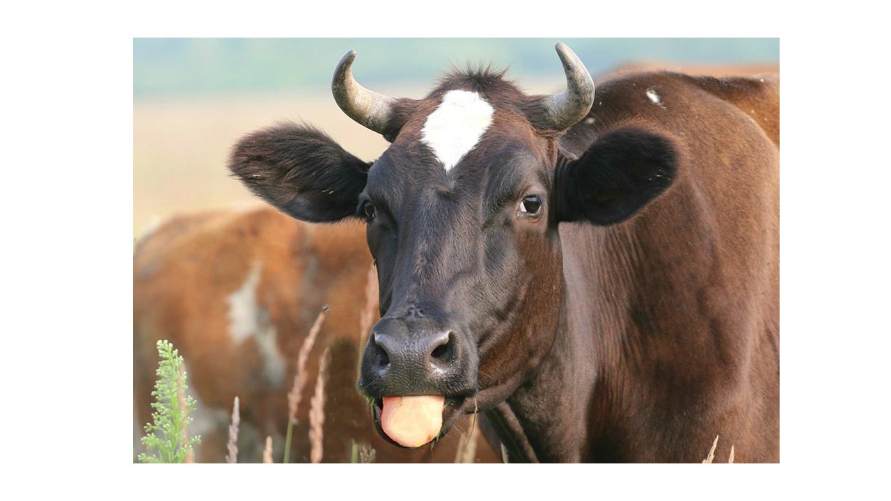
Actinobacillosis is a disease caused by the bacterium, Actinobacillus spp. There are over 20 known species of Actinobacillus however, five of them are known to cause disease in animals. Each of the Actinobacillus spp exhibits different signs in various species of animals. Our focus today will be on Actinobacillus lignieresii, which causes Wooden Tongue mainly in cattle. The disease can also be seen in sheep, horses, pigs and dogs. The causal organism Actinobacillus spp, is a commensal which resides in the upper digestive tract of animals. Infection occurs usually after an injury to the inner lining of the buccal cavity (mouth). During grazing, ruminants sometimes get cuts from sharp grasses or objects. This allows the bacterium to have access to the tissues of the buccal cavity, leading to the disease. The disease can spread from the tissues surrounding the mouth to other regions.  Signs of the disease usually seen include:

  1. Sudden swelling around the lower jaw, face, and sometimes the neck and chest regions with pus discharge
  2. Swollen and hardened tongue which is painful to touch. This is usually observed in cattle
  3. Sores on the tongue
  4. Excessive drooling with protruding tongue

Actinobacillosis can be diagnosed by culture, ELISA or PCR. In severe cases, surgical therapy is considered. Topical and broad-spectrum systemic antibiotics have proven effective in the treatment of wooden tongue. Consult your veterinarian once you see similar signs for assistance.

HAEMONCHOSIS

HAEMONCHOSIS 1280 720 Kingsley Emmanuel Bentum

Photo Credit: http://www.sheep101.info/201/parasite.html

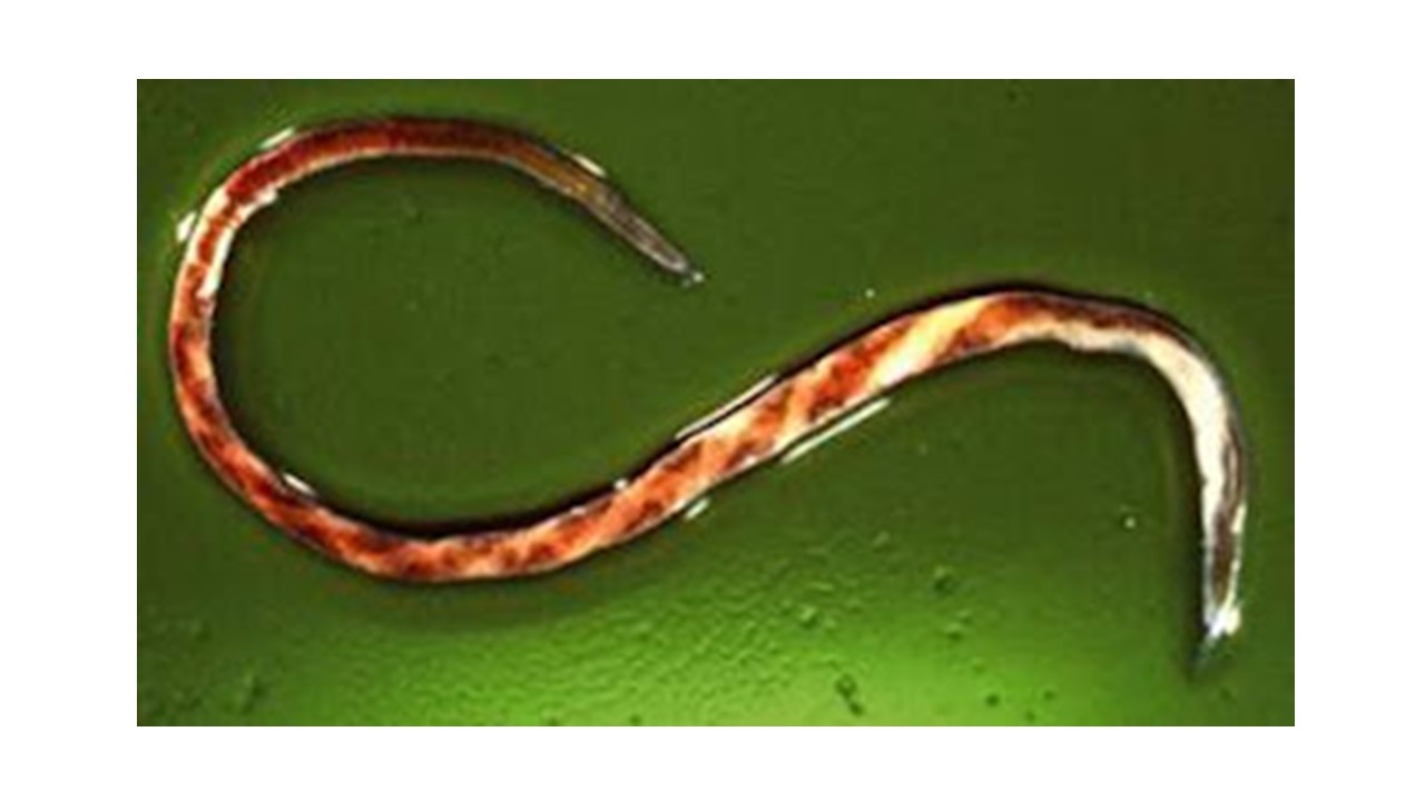
Amid the several parasitic infections that plague ruminants, Haemonchus spp has been recognized as one of the many parasites that cause severe damages to the digestive system. This blood-sucking worm is responsible for a lot of losses in cattle, sheep and goat production.

A parasite generally is any organism, that lives in or on another (host) and harms or offers no advantage to the other organism (host).

This worm also commonly referred to as Barber’s Pole worm resides in the abomasum of ruminants (fourth or last chamber of the ruminant’s stomach).  Haemonchus spp has piercing mouthparts that causes extensive damage to the walls of the abomasum. An adult worm is capable of sucking about 0.05mls of blood daily from an affected animal. Hence an animal infected with about 2000 worms will have 100mls of blood loss daily.

There are various species of the blood parasite affecting various ruminant types. Some of the common species of Haemonchus include Haemonchus contortus, Haemonchus similis and Haemonchus placei. There are few records of cross-transmission of the Haemonchus spp between small ruminants and cattle.  Haemonchosis has remained a threat to ruminant production especially in the tropical, subtropical and warm temperate regions of the world, where environmental conditions favour the free-living stages of the parasite.

CLINICAL SIGNS & DIAGNOSIS

In acute conditions, anaemia, agalactia in ewes, submandibular oedema or bottle jaw and death due to blood loss are among the common clinical signs. When the condition becomes chronic, there is progressive weight loss and general weakness. Diagnosis is based on presenting clinical signs, grazing history as well as worm egg count from the laboratory.

TREATMENT & PREVENTION

Consult your veterinarian once you notice similar signs on your farm. Having a good management program to control parasitic infection on your farm is important.

="ringworm in pets"

RINGWORM IN PETS

RINGWORM IN PETS 494 363 Ekua Esuon Thompson

Photo Credit: © 2022 Merck & Co., Inc.

The name Ringworm may suggest an infection caused by a worm; however, this condition isn’t caused by a worm at all, but a fungus, “Microsporum canis” which can cause a generalized skin infection.  It is often associated with severe hair or fur loss and creates a ‘worm-like’ or circular rash at the site of infection, hence the name Ringworm. This highly contagious disease easily spreads to other animals and humans as well.

CAUSE AND SOURCE OF INFECTION

Most often, the source of infection is through direct contact with an infected pet or through the sharing of contaminated bedding, kennels (especially in shelters and breeding houses where dogs comingle and crowd together), toys, rugs, feeding bowls, etc. The fungal spores can remain dormant and only grow under favourable environmental conditions. Animals that like digging and playing in the soil also expose themselves to the fungus.  It is worth noting that, dogs (especially puppies less than a year old) are more prone to ringworm infection. Similarly, malnourished, immunocompromised, and stressed dogs are also at a greater risk of infection.

SIGNS AND SYMPTOMS

Key symptoms of ringworm in dogs include:

  • Skin lesions that typically appear on the head, ears, paws and limbs.
  • Patchy, crusted, scaly and circular bald spots that sometimes look red in the center and may be itchy.
  • In mild cases, there may be just a few broken hairs, whilst in severe cases it spreads over most of a dog’s body.
  • Toenails may be brittle and easily broken.

NB: It’s also possible for a pet to carry the fungus and not show any symptoms whatsoever.

DIAGNOSING RINGWORM

It is necessary that you see your vet for an accurate diagnosis if your pet is showing any signs of a skin problem as the infection can potentially spread over your pet’s body and infect other animals and people. Diagnostic options available to a veterinarian may include using an ultraviolet light (Wood Lamp examination) to see the extent of the infection, or examining a fungal culture taken from the affected area.

TREATMENT AND PREVENTION

Treatment of ringworm depends on the severity of the infection. A veterinarian may prescribe a medicated shampoo or ointment that kills fungi. In some cases, oral medications are necessary, but usually for long periods. It is important to treat your dog for as long as recommended by your veterinarian. Unfortunately, there is no guarantee that reinfection won’t occur.

If your veterinarian has diagnosed your dog with ringworm, he or she will explain what you must do to prevent the fungus from spreading to other pets and human members of the household. But keep in mind that if you have other pets, it’s likely that most of them have been exposed as well. Your veterinarian may recommend that you do the following:

  • Bathe all pets in the household with a medicated rinse or shampoo.
  • Wash the infected animals’ bedding and toys with a disinfectant that kills ringworm spores.
  • Discard items that are impossible to thoroughly disinfect (e.g., carpeted cat trees)
  • Frequently vacuum to rid the house of infected hairs and skin cells as the fungus can survive on hair and skin that your dog sheds.
  • Thoroughly wash your hands after you bathe or touch your pet.
="image or psittacosis

PSITTACOSIS

PSITTACOSIS 1557 1603 Ekua Esuon Thompson

It is a highly contagious disease, with a mortality rate of about 50% among birds.  Several species of birds (over 465 species) can be affected by this disease, however, birds of the Parrot family or Psittacines are the most affected, hence the name psittacosis. It is also referred to as Parrot Fever.Turkeys and ducks are more susceptible to this disease compared to chickens. This is also a zoonotic disease; this means that humans are at risk of contracting this disease.

Psittacosis is caused by the bacterium, Chlamydia psittaci. There are several subtypes (strains) of Chlamydia psittaci. Some of these strains are known to cause severe infections in humans. Healthy birds or humans can get infected through inhalation or via a  faecal-oral route from infected birds (they could be sick/ diseased birds or carriers; they do not show signs of the disease but can spread the bacterium). Other sources of infection include contact with infected arthropods and contaminated environment (feed, water, toys, perch). Mother to chicks (vertical transmission) transmission is possible.

Clinical Signs include: weight loss, swollen and watery eyes, ocular and nasal discharges, ruffled feathers, yellowish or green droppings, breathlessness. Therapy is based on laboratory findings and presenting clinical signs. Treatment can last from about two weeks to a month. Birds who recover remain carriers of the bacterium and can potentially spread to other birds in the future.

It is important to protect yourself when handling a bird infected with psittacosis. Put on protective gear, mask and gloves when cleaning their cages and it is recommended to disinfect or wash your hands and clothing afterwards. All new birds (pets or poultry) should be quarantined, for a minimum of 14 days to check for possible clinical signs of this important disease.

="canine ehrlichiosis"

CANINE EHRLICHIOSIS

CANINE EHRLICHIOSIS 2560 1700 Ekua Esuon Thompson

Canine Ehrlichiosis is bacterial disease which occurs worldwide, especially in tropical countries. The disease is transmitted via the brown dog tick (Rhipicephalus sanguineus). There have been few reports of transmission through blood transfusion. Several species of Ehrlichia are known to infect dogs including Ehrlichia ewigii, Ehrlichia equi and Ehrlichia platys. However, Ehrlichia canis causes the most common and severe form of disease in dogs.

TRANSMISSION

An infected brown dog tick transmits the adult stage or nymphs of Ehrlichia to a new host during feeding (blood meal). The bacterium invades and multiplies in the hosts’ monocytes, lymphocytes and reticuloendothelial cells (immune response cells). The infection could be acute, beginning after an incubation period of averagely two weeks. The disease becomes chronic after the organism persists, for more than three months.

CLINICAL SIGNS

Signs of disease include fever, weight loss, anemia, abdominal pain, shifting lameness and seizures.

DIAGNOSIS, PREVENTION & CONTROL

Laboratory blood tests are usually used to confirm Ehrlichiosis. Your pet will most likely be placed on a long-term therapy, usually of about four weeks.

It is important to seek medical care as soon as you notice your pet exhibiting similar signs as mentioned above. The most effective way to prevent this disease is the effective control of ticks.  Discuss with your Vet the available tick control options, and the one that is best suited for your pet.

="image of ecto-parasites in pets: ticks & fleas".

ECTO-PARASITES IN PETS: TICKS &FLEAS

ECTO-PARASITES IN PETS: TICKS &FLEAS 466 269 Ekua Esuon Thompson

Photo Credit: © 2022 by Advanced Care Veterinary

It is our desire to see our cherished pets always healthy and happy. One of the most worrying issues in taking care of our pets are ectoparasites. They just seem to always be around no matter what we do. Sometimes we resort to desperate measures in our quest to kill these irritating organisms. These parasites are more powerful than they look.

Below are some facts about ticks and fleas.

  1. Fleas are dark brown, wingless insects, with laterally compressed bodies which have a glossy surface, allowing easy movement through hairs and feathers of their host. The third pair of legs are much longer than the others, an adaptation for leaping on and off their hosts.
  2. In fleas, both sexes are blood suckers but only the adults are parasitic and the life span for fleas is averagely 1-2 years.
  • The flea spp in Dogs and Cats act as an intermediate host for tapeworm infection in dogs. This tapeworm known as Dipylidium caninum is zoonotic (i.e., can infect humans).
  • Ticks belong to a group called Arachnids (eight-legged organisms). There are generally two kinds of ticks, the hard tick (because they have a hard or rigid covering on the dorsal surface of the tick, called the scutum) and the soft tick (they lack the hard chitinous scutum).
  • All stages of the life cycle of the tick suck blood, especially the female tick. One female tick lays an average of 2500-3000 eggs at a time.
  • Ticks cannot jump or fly so they use the blades of grasses and other vegetation to elevate themselves and grasp onto moving animals (this is known as questing).
  • Ticks aside feeding on blood also transmit diseases to both animals and humans such as Babesiosis, Anaplasmosis, Ehrlichiosis and Lyme disease.

BAD PRACTICES TO AVOID WHEN YOU SEE THESE PARASITES ON YOUR PET

  1. Picking ticks with your bare hands and squashing or crushing them. This is because ticks can habour several zoonotic pathogens and squashing them release these pathogens into the environment.
  2. Forcefully picking ticks off pets can lead to some of the mouth parts of the parasites remaining buried in the skin of your pet. This can cause irritation and inflammation of the skin.
  3. Using unknown products and chemicals with the aim of killing ticks and fleas. Some of these products may be harmful to your pet, your household, and the environment.

PREVENTION/CONTROL

  1. Bath your pet regularly
  • Clean your pet’s kennel or cage as frequently as possible
  • Mow your lawn frequently
  • Use of flea combs where applicable
  • There are various products and chemicals available, both topical and oral, which can be used to kill the parasites. Talk to your vet or visit a pet shop available in your area and be assisted in choosing the best product suitable for your pet
="image of dog with kennel cough

KENNEL COUGH

KENNEL COUGH 555 396 Ekua Esuon Thompson

Photo Credit: Copyright © 2022 · Top Notch Pet Care

Kennel cough is a term loosely used to describe a complex of respiratory infections (both viral and bacterial), that cause inflammation of a dog’s larynx (voice box) and trachea (windpipe). It’s a form of bronchitis (inflammation of the bronchi) and is similar to a chest cold in humans. Dogs are more prone to Kennel Cough.  Though it usually clears up on its own, kennel cough is highly contagious and a source of worry to many owners.

Cause

As Kennel Cough is a respiratory infection, it is spread mainly via aerosols in the air. Dogs prone to this condition are those:

  • In breeding homes. The spread is more rapid when the area is enclosed with poor air circulation.
  • Who have frequent contact with other dogs, in open environments especially where the environment is wet and humid.
  • Who have contact with contaminated materials such as eating and drinking bowls, etc.
  • Who have not been vaccinated against Parainfluenza and Bordetella which are two of the main causes of kennel cough.

Symptoms/Signs

  • A persistent dry cough with a “honking” sound. In most cases, pets appear healthy except for the cough.
  • Gagging, Coughing up white foamy phlegm
  • Fever normally accompanied with nasal discharge

Treatment and Prevention

  • Dogs with kennel cough should be isolated from other dogs. A humidifier, vaporizer or steam from a shower can provide relief for irritated breathing passages.
  • Avoid exposing your dog to cigarette smoke or other noxious irritating fumes.
  • A cough suppressant and antibiotics may be prescribed. Consult your veterinarian for the right drug suited for your pet.
  • If your dog pulls against her collar while being walked, replace it with a harness until the coughing subsides.
  • Supportive care is very important. Be sure your dog is eating, drinking and in a stress-free environment.
  • Vaccinations are also available for several of the agents known to be involved in kennel cough including parainfluenza, bordetella and adenovirus 2.  Consult your veterinarian on which ones are recommended for the environment you find yourself in and how often.
  • Vaccinations aren’t useful if a dog has already caught the infection.

Kennel Cough Recovery

In most cases, the signs gradually decrease and disappear after three weeks. Young puppies, elderly dogs and other immunocompromised animals may take up to six weeks or more to recover. Animals may remain infectious for long periods of time even after the symptoms have cleared up.

When to Consult Your Veterinarian

If your dog has nasal discharge, is breathing rapidly, refuses to eat or seems lethargic, take it to the veterinarian right away and immediately isolate it from all other dogs. Serious cases of this disease can lead to pneumonia if left untreated.

="image of ferocious dog. Help Save a life, Vaccinate your pet. Rabies

HELP SAVE A LIFE, VACCINATE YOUR PET

HELP SAVE A LIFE, VACCINATE YOUR PET 589 399 Ekua Esuon Thompson

Photo Credit: https://www.flickr.com/photos/dgoomany/6338142380

Rabies is the most common zoonotic disease spoken of when issues relating to pet health comes up. This disease has killed thousands and continues to do so every year. In Africa alone, on average about 59,000 people die annually because of this disease. Ghana is no exception, records of several dog bites with confirmed rabies cases have been realized in many regions of the country. The sad thing is that these deaths could easily have been prevented by vaccinating our pets.

TRANSMISSION

Rabies is a viral infection that affects mammals (warm blooded animals whose females have milk secreting glands to feed their young); both domesticated and wild. Dogs, cats, bats, and rats are examples of animals prone to this disease. Humans are also mammals and therefore can be infected with the virus. The disease is transmitted through the saliva of an infected animal usually by bites or scratches from the animal. In special conditions such as experimental laboratories and bat caves with high concentrations of the virus, one can get infected by inhalation.

The virus then travels to the brain which is its main site of replication (predilection site). The virus multiplies and is released in high concentrations in the salivary gland. How long the virus takes to get to the brain and for replication to take place is dependent on the proximity of the wound to the brain. Duration can be as short as 3-4 days to as long as months or even years. For the disease to manifest itself, it is also dependent on the amount of virus transmitted, viral strain, site of bite, nature of the wound and host immunity.

In animals, two forms of rabies are observed depending on the site of brain affected by the virus. There is the furious form which is the most popular and widely observed and the dumb/paralytic form.

SIGNS OF RABIES IN ANIMALS

– Inability to swallow or severe drooling

– Facial paralysis

– Highly aggressive

– Fear of exposure to light

– Difficulty in breathing

– Lameness

SIGNS OF RABIES IN HUMANS

– Headaches

– Fear of water

– Fear of light

– Difficulty in swallowing and drinking

– Hallucinations

– Anxiety

The danger with this disease is that once signs begin to show, there is nothing that can be done to treat the disease. Death comes knocking on the door of the victim.

Rabies is diagnosed in the laboratory by taking brain tissues from infected animals. Recent studies have identified other samples aside from the brain to test for the virus. Tests include RT-PCR, ELISA and immunofluorescent tests. 

WHAT TO DO WHEN BITTEN BY A DOG

– Wash the site of bite with running water and soap for a minimum of 15 minutes

– Disinfect the wound with alcohol or iodine

– Go to the nearest hospital immediately

WHAT HAPPENS TO THE DOG THAT BIT AN INDIVIDUAL

– Report the case of dog bite to the veterinarian in your vicinity. The Veterinarian will quarantine the dog and examine it to ensure that it is not a threat to you and to everyone in the town.

– Don’t make an attempt to harm or kill the dog.

PREVENTION

– Vaccinate your dog or cat at 3 months of age at the veterinary hospital close to you

– Avoid letting your pets stray around

– Provide your pet with food and water daily – Keep your pet on a leash always when going out for walks

="image of heart infested with worms. Canine heartworm disease"

CANINE HEARTWORM DISEASE

CANINE HEARTWORM DISEASE 1933 1363 Ekua Esuon Thompson

Photo Credit: https://journals.asm.org/doi/10.1128/CMR.00012-12

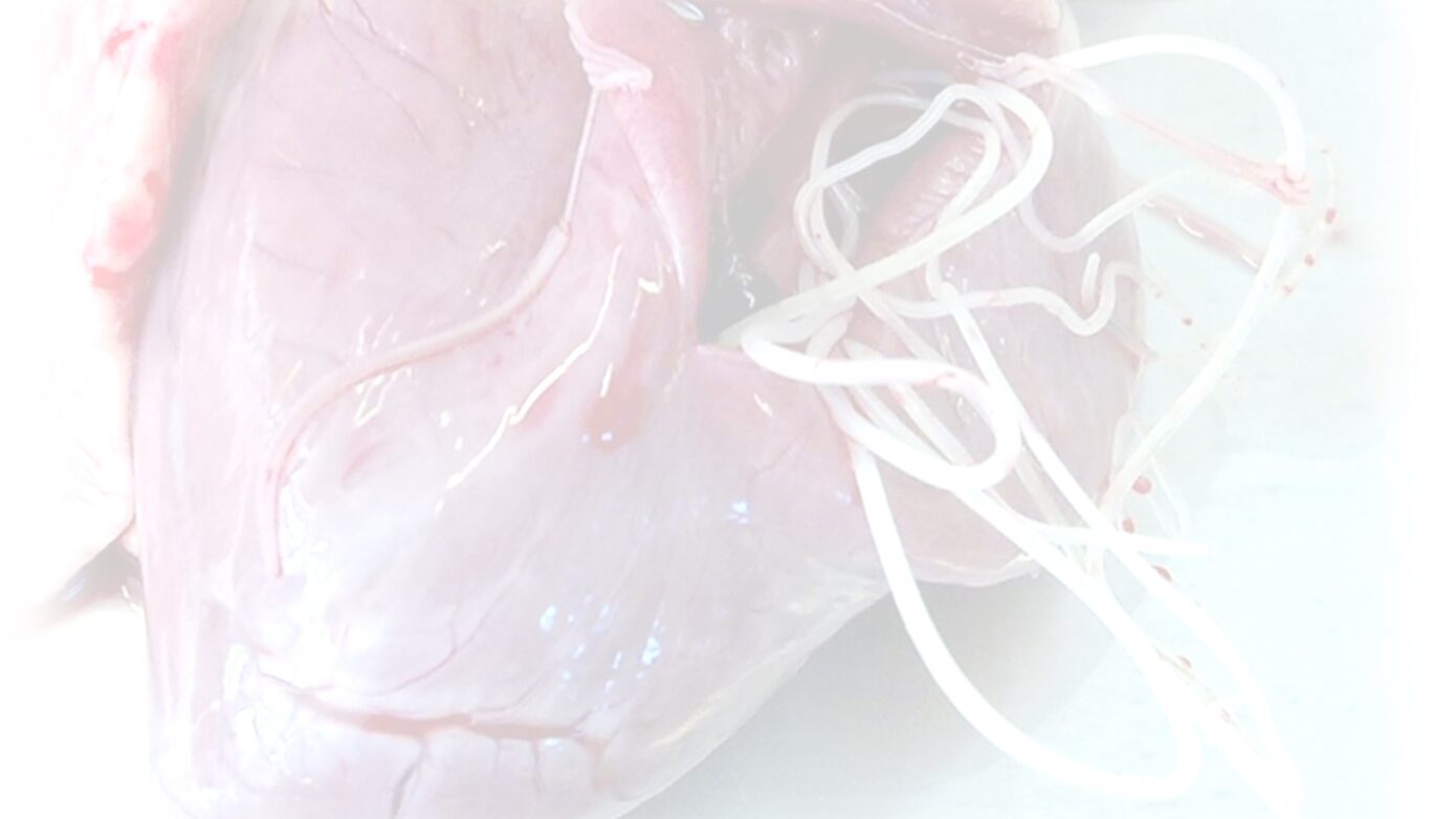
Canine heartworm disease is a potentially deadly disease caused by the worm Dirofilaria immitis. This parasitic worm lives in the heart and lungs (pulmonary arteries) of an infected dog. The worm travels through the bloodstream, harming arteries and vital organs as they go. Ultimately, it completes its journey to the vessels of the lung and the heart chamber about six months after the initial infection. Several hundred worms can live in one dog for as long as five to seven years. Dogs are natural hosts for heartworm infection however, other animals can be infected such as cats, ferrets and even humans (though it has not been recognized to be a major health issue). The disease is fatal but also preventable.

TRANSMISSION OF HEARTWORM DISEASE

  • The source of this disease is a mosquito bite transmitting larvae of the worm Dirofilaria immitis. It has been reported that there are at least 70 species of mosquitoes that can transmit the disease including Aedes, Anopheles and Mansonia species. Humans and other mammals are accidental hosts of the worm. You do not get infected by having contact with your pet.
  • An animal must carry at least two heartworms (a male and a female) in order for female heartworms to reproduce.
  • Females produce “babies” called “microfilaria” which are shed into an animal’s bloodstream but are not capable of directly causing the heartworm disease as they need further development in the mosquito. The microfilariae must be taken up by biting mosquitoes and get transformed into infective larvae over a two-week period inside the mosquito.
  • When a mosquito next bites a susceptible animal, the infective larvae enter the tissues and begin a migration into the blood vessels.
  • Heartworms enter an animal’s bloodstream as tiny, invisible larvae, but can reach lengths of more than twelve inches at maturity.

SIGNS AND SYMPTOMS

Severity of signs is dependent on the worm load, the immune status of the animal and the duration of the infection. Some dogs exhibit no symptoms at all until late stages of infection while others may show signs such as: labored breathing, coughing, vomiting, weight loss, listlessness, and fatigue after even a moderate exercise.

DIAGNOSIS AND TREATMENT

Heartworm disease is diagnosed by examination, radiographs or ultrasound, and a blood test. After diagnosis, a thorough examination of the infected dog should be conducted to evaluate the best course of treatment and the potential risks involved. There are various drugs, both topical and intravenous to cater for the infection.  All treatment protocols will require at least several weeks of exercise restriction during and after treatment. All dogs should be routinely screened with a blood test for heartworm either annually or before being placed on a new prescription for the prevention of heartworm.

PREVENTING HEARTWORM

Heartworm is easily preventable with an inexpensive, chewable pill or topical medication available at a vet’s prescription. The drugs are usually administered monthly and can be given to dogs under 6 months of age without a blood test. Older animals must be screened for the disease prior to starting medication. Keep your dog on preventive medication all year long. Many of these medications also prevent other intestinal parasites. The use of adulticides to also kill adult mosquitoes that spread and transmit these worms is also recommended. If you notice that your dog’s energy has decreased, he seems ill, or he’s exhibiting any of the general symptoms described above, contact your veterinarian immediately.