Ruminants

SHIPPING FEVER

SHIPPING FEVER 1280 720 Kingsley Emmanuel Bentum

Photo Credit: @2020 All rights observed by Orissa

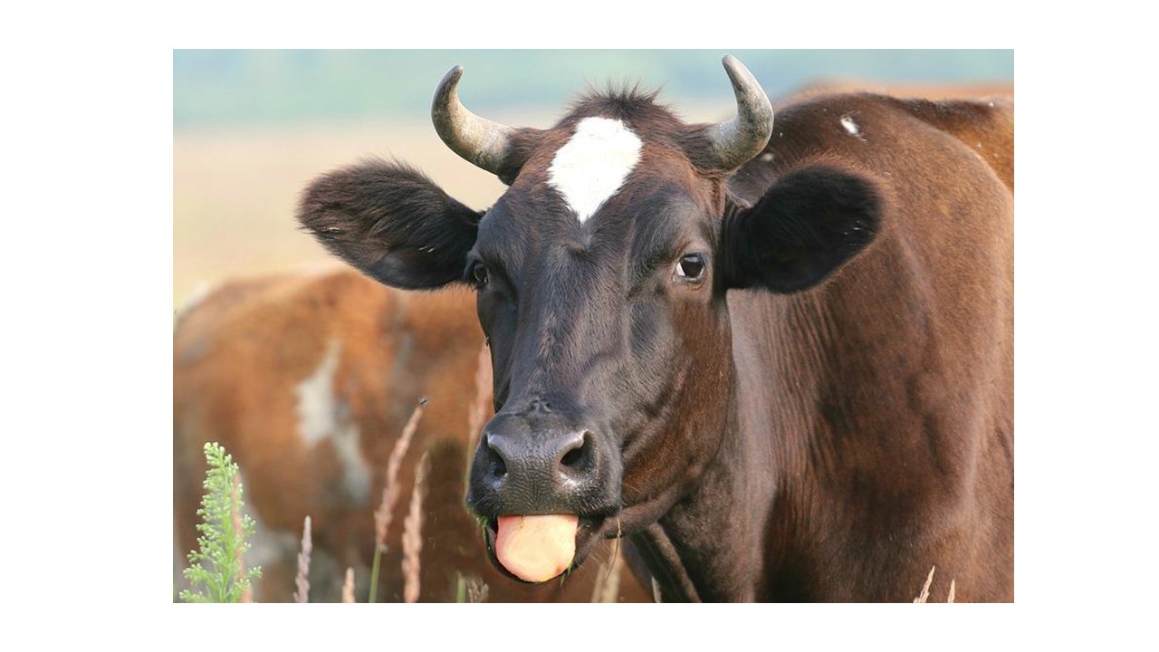
Shipping Fever also known as Bovine Respiratory Disease Complex is a bacterial disease of cattle usually associated with stress involved in movement. The disease affects the upper respiratory system and is presented with signs such as laboured or rapid breathing, nasal discharges, coughing, anorexia and depression. 

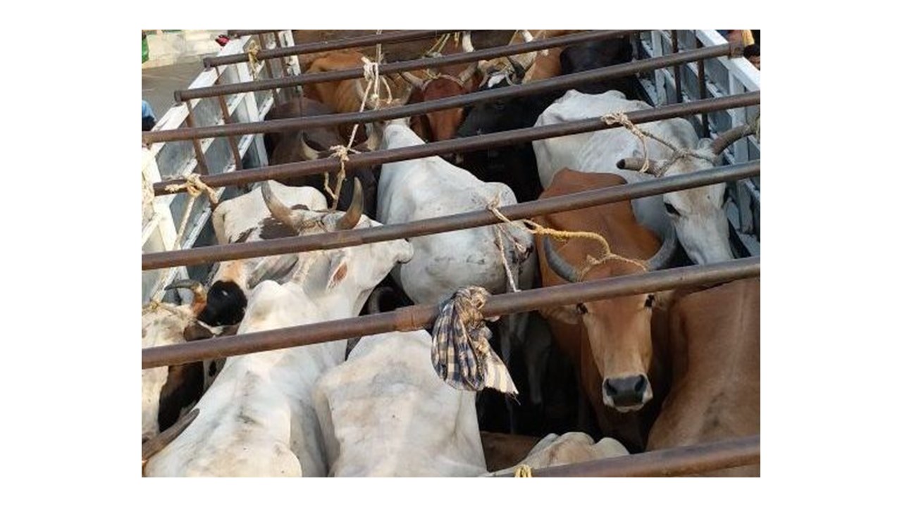
Stress factors associated with movement of animals such as long journeys with little or no rest, starvation, overheating due to poor ventilation in trucks, dust and harsh weather conditions weaken the immune systems of the cattle, and predispose the animals to this disease. Shipping fever is caused by the bacterium Mannheimia haemolytica. It is often complicated by other bacteria; Pasteurella multocida and Histophilus somni.  Already sick or weak animals  and recently weaned calves have a higher chance of contracting the disease due to their compromised and fragile immune systems. While Shipping Fever is usually not fatal, it affects productivity greatly as several animals come down with the infection within a short time.

Elimination of all possible stressors  is the first step for recovery in cattle. Provision of adequate water, open space with fresh air and food are few examples to take note of. Some antibiotics may be prescribed by your veterinarian depending on the implicating bacterium. Vitamin and mineral supplements may be added to the therapy as animals may be weak and would not have enough energy to move about.

Preventive measures for Shipping Fever should include providing resting periods for animals being transported especially during long journeys. The rest period should include provision of food and water for animals. Trucks transporting animals should not be overloaded and must be roomy enough to allow them to breathe and move. It is not advisable to keep sick and healthy animals in the same truck. Calves should be weaned at least 2-3 weeks before transportation. At various animal markets, shelters should be created to protect them from the harsh environmental conditions.

Vaccines for Shipping Fever are available and should be given at least 2–3 weeks prior to transportation.  Vaccination can be repeated when animals are introduced into feedlot.

HAEMONCHOSIS IN RUMINANTS

HAEMONCHOSIS IN RUMINANTS 1280 720 Kingsley Emmanuel Bentum

Photo Credit: Adduci, Isabella, et al. “Haemonchosis in Sheep and Goats, Control Strategies and Development of Vaccines against Haemonchus contortus.” Animals 12.18 (2022): 2339.

Amid the several parasitic infections that plague ruminants, Haemonchus spp has been recognized as one of the many parasites that cause severe damages to the digestive system. This blood-sucking worm is responsible for a lot of losses in cattle, sheep and goat production.

A parasite generally is any organism, that lives in or on another (host) and harms or offers no advantage to the other organism (host).

This worm also commonly referred to as Barber’s Pole worm resides in the abomasum of ruminants (fourth or last chamber of the ruminant’s stomach).  Haemonchus spp has piercing mouthparts that causes extensive damage to the walls of the abomasum. An adult worm is capable of sucking about 0.05mls of blood daily from an affected animal. Hence an animal infected with about 2000 worms will have 100mls of blood loss daily.

There are various species of the blood parasite affecting various ruminant types. Some of the common species of Haemonchus include Haemonchus contortus, Haemonchus similis and Haemonchus placei. There are few records of cross-transmission of the Haemonchus spp between small ruminants and cattle.  Haemonchosis has remained a threat to ruminant production especially in the tropical, subtropical and warm temperate regions of the world, where environmental conditions favour the free-living stages of the parasite.

CLINICAL SIGNS & DIAGNOSIS

In acute conditions, anaemia, agalactia in ewes, submandibular oedema or bottle jaw and death due to blood loss are among the common clinical signs. When the condition becomes chronic, there is progressive weight loss and general weakness. Diagnosis is based on presenting clinical signs, grazing history as well as worm egg count from the laboratory.

TREATMENT & PREVENTION

Consult your veterinarian once you notice similar signs on your farm. Having a good management program to control parasitic infection on your farm is important.

DIARRHOEA IN NEWLY BORN RUMINANTS CAUSED BY E.coli

DIARRHOEA IN NEWLY BORN RUMINANTS CAUSED BY E.coli 1280 720 Kingsley Emmanuel Bentum

Diarrhoea in newly born ruminants caused by Escherichia coli (E.coli) remains as one of the common conditions of newborns which sometimes leads to death.

E. coli, is a bacterium which naturally resides in the intestines of living organisms and aid in digestion. There are however some strains of E. coli (Enterotoxigenic E. coli and Enteropathogenic E. coli) that cause severe infection especially in the early lives (normally within the first week) of calves and lambs. The infection is sometimes complicated with other pathogens such as Clostridium perfringens and Rota virus.

Enterotoxigenic E. coli release enterotoxins which disrupts the osmotic balance of the gut cells, leading to excess fluid secretion into the lumen of the gut, resulting in diarrhoea.  

Enteropathogenic E. coli affect the intestinal lining, leading to ulcers and erosions of the intestinal walls. Some of these strains of E. coli release toxins (verotoxin) which enhance the erosions of the intestinal mucosa leading to hemorrhages.

Clinical Signs

Diarrhoea (very clear watery stool or bloody)

– Dehydration

– Death

– Lethargy

– Fever

Treatment and Prevention

– Practice good hygiene especially in delivery pens

– Ensure young animals get colostrum especially during the first week of life. Kids (baby goats)  are however resistant to this infection.

Affected animals are usually given supportive therapy with antibiotics after sensitivity testing has been carried out.

DETERMINE THE AGE OF YOUR SHEEP OR GOAT USING ITS’ TEETH

DETERMINE THE AGE OF YOUR SHEEP OR GOAT USING ITS’ TEETH 1280 720 Kingsley Emmanuel Bentum

Today’s article is a very useful tool in guiding your decision making during buying new animals both for breeding and production purposes. Veterinarians generally use this technique also, to guess the approximate age of an animal where data or acquisition history of the animal may be deficient. In the absence of any defect or accidents, dentition of ruminants has proven and remained one of the popular and easy means to determine their age.

An adult sheep or goat has a total of 32 teeth. There are four pairs of incisors and twelve molars on the lower jaw whilst twelve molars and a dental pad are found on the upper jaw. Lambs and kids have temporary/milk teeth (incisors) which will eventually fall out and be replaced by permanent teeth.

The incisors therefore play a major role in enabling us to approximate the age of a sheep or goat (I I I I I I I I). When your ruminant is about a year old, the middle pair of incisors get replaced by a pair of permanent incisors (I I I I I  I I I).  After another year (2 years old), they have an additional pair of incisors replaced by permanent teeth (I l I  I I  I l  I). By their fourth year, all four incisors would have been replaced by permanent teeth (I I I I I I I I).

As they animals age (beyond 5 years), the teeth begin to space out, and then break or fall out. A sheep whose teeth are all fallen out (approximately 10 years old) is called a ‘’gummer”/” gummy’’.

Picture credit:Royal Veterinary CollegeAttribution-Noncommercial 4.0 International

BRUCELLOSIS (MEDITERRANEAN FEVER/ UNDULANT FEVER) IN SMALL RUMINANTS

BRUCELLOSIS (MEDITERRANEAN FEVER/ UNDULANT FEVER) IN SMALL RUMINANTS 1280 720 Kingsley Emmanuel Bentum

Brucellosis is a highly infectious disease that has the potential of being transferred from animals to humans (zoonotic). It makes it a disease of great public health significance with economic impact.

ETIOLOGY / CAUSATIVE AGENT

It is caused by the bacterium Brucella spp. Brucella has different species including Brucella abortus, Brucella canis, Brucella suis  and Brucella melitensis. These species affect several animals such as cattle, goats, sheep, pigs, dogs, horses and camels. Recent scientific discoveries have identified strains of brucella in red foxes and certain marine animals. Brucella melitensis is the main strain that affects sheep and goats and also has the potential of infecting humans.

MODE of INFECTION

Brucella spp is spread through contact with contaminated birth or uterine fluids, milk and semen from infected animals. Other means of contracting the disease include cuts or scratches through the mucous membranes. 

CLINICAL SIGNS

These may include abortions during the late stages of pregnancy, retained placentas, still births, weak offspring, swollen or inflamed testicles (orchitis) and arthritis. In human, symptoms may include fever, headaches, muscle, and joint pains.

PREVENTION and CONTROL

Brucellosis is difficult to be treated and hence no practical treatment protocol exist in animals. Testing of animals and the elimination of positive reactors is the recommended protocol for control and prevention of the disease. It is important to purchase animals from credible sources and test breeding stock for brucellosis before mating them. After birth discharges should be properly disposed off to reduce spread of infection

ACTINOBACILLOSIS (Wooden Tongue)

ACTINOBACILLOSIS (Wooden Tongue) 1280 720 Kingsley Emmanuel Bentum

Actinobacillosis is a disease caused by the bacterium, Actinobacillus spp. There are over 20 known species of Actinobacillus however, five of them are known to cause disease in animals. Each of the Actinobacillus spp exhibits different signs in various species of animals. Our focus today will be on Actinobacillus lignieresii, which causes Wooden Tongue mainly in cattle. The disease can also be seen in sheep, horses, pigs and dogs. The causal organism Actinobacillus spp, is a commensal which resides in the upper digestive tract of animals. Infection occurs usually after an injury to the inner lining of the buccal cavity (mouth). During grazing, ruminants sometimes get cuts from sharp grasses or objects. This allows the bacterium to have access to the tissues of the buccal cavity, leading to the disease. The disease can spread from the tissues surrounding the mouth to other regions.  Signs of the disease usually seen include:

  1. Sudden swelling around the lower jaw, face, and sometimes the neck and chest regions with pus discharge
  2. Swollen and hardened tongue which is painful to touch. This is usually observed in cattle
  3. Sores on the tongue
  4. Excessive drooling with protruding tongue

Actinobacillosis can be diagnosed by culture, ELISA or PCR. In severe cases, surgical therapy is considered. Topical and broad-spectrum systemic antibiotics have proven effective in the treatment of wooden tongue. Consult your veterinarian once you see similar signs for assistance.