Ruminant

Sheep and Goats.

BLOAT/ RUMINAL TYMPANY

BLOAT/ RUMINAL TYMPANY 1280 720 Kingsley Emmanuel Bentum

Bloat is an uncomfortable condition in ruminants marked by an over distension of the rumen (the first of the four divisions of stomach in ruminants). There are microbes which are naturally found in the rumen and aid in the fermentation process of feed. Gas naturally produced due to this process is expelled by eructation or burping. Bloat occurs when there is any form of hindrance to the normal release of gas from the rumen.

TYPES OF BLOAT

Free-Gas Bloat: This is a less common type, and it occurs due to the oesophagus being blocked by a foreign object such as a lump of feed. There are other medical conditions such as tetanus, tumors and hypocalcaemia (reduced calcium levels in the blood) which can affect the movement of the oesophagus, leading to this condition.

Frothy Bloat: This is the most common type of bloat. It occurs usually during the onset of the rainy season when there is a lot of fresh highly proteinous forage such as legumes. These plants are rapidly digested leading to the formation of a layer of entrapped gasses in the form of foam, which makes it difficult to be released from the rumen.

CLINICAL SIGNS

  • A highly distended left abdomen
  • Sudden death
  • Difficulty in breathing (dyspnea)
  • Death can occur within 4 hours after signs of bloat begin to show due to an impairment of normal respiration.

DIAGNOSIS

Diagnosis is usually based on history and physical examination. The use of the stomach tube is useful in distinguishing between free-gas and frothy bloat

TREATMENT and PREVENTION

  1. A stomach tube or Trocar and Cannula is used to release excess gas from the rumen. In life threatening cases a surgical procedure may be needed.
  2. Antifoaming products such as Poloxalene, vegetable and mineral oils have proven to be effective.

Avoid grazing animals on high-risk pastures. Diet of animals fed in stalls should contain a balance of grains and roughage. You may consider providing your farm animals antifoaming agents especially during the rainy season when there is a lot of lush vegetation. Ensure that feed or pastures are always free from foreign materials such as plastics and stones.

ACTINOBACILLOSIS (Wooden Tongue)

ACTINOBACILLOSIS (Wooden Tongue) 1280 720 Kingsley Emmanuel Bentum

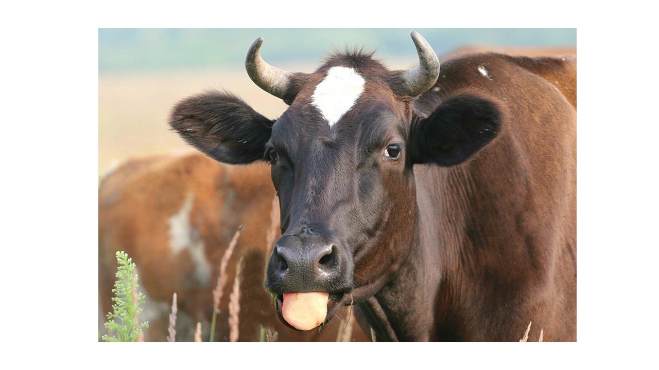
Actinobacillosis is a disease caused by the bacterium, Actinobacillus spp. There are over 20 known species of Actinobacillus however, five of them are known to cause disease in animals. Each of the Actinobacillus spp exhibits different signs in various species of animals. Our focus today will be on Actinobacillus lignieresii, which causes Wooden Tongue mainly in cattle. The disease can also be seen in sheep, horses, pigs and dogs. The causal organism Actinobacillus spp, is a commensal which resides in the upper digestive tract of animals. Infection occurs usually after an injury to the inner lining of the buccal cavity (mouth). During grazing, ruminants sometimes get cuts from sharp grasses or objects. This allows the bacterium to have access to the tissues of the buccal cavity, leading to the disease. The disease can spread from the tissues surrounding the mouth to other regions.  Signs of the disease usually seen include:

  1. Sudden swelling around the lower jaw, face, and sometimes the neck and chest regions with pus discharge
  2. Swollen and hardened tongue which is painful to touch. This is usually observed in cattle
  3. Sores on the tongue
  4. Excessive drooling with protruding tongue

Actinobacillosis can be diagnosed by culture, ELISA or PCR. In severe cases, surgical therapy is considered. Topical and broad-spectrum systemic antibiotics have proven effective in the treatment of wooden tongue. Consult your veterinarian once you see similar signs for assistance.

HAEMONCHOSIS

HAEMONCHOSIS 1280 720 Kingsley Emmanuel Bentum

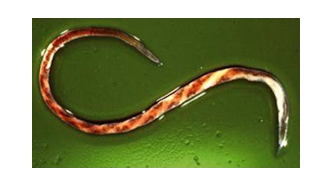
Photo Credit: http://www.sheep101.info/201/parasite.html

Amid the several parasitic infections that plague ruminants, Haemonchus spp has been recognized as one of the many parasites that cause severe damages to the digestive system. This blood-sucking worm is responsible for a lot of losses in cattle, sheep and goat production.

A parasite generally is any organism, that lives in or on another (host) and harms or offers no advantage to the other organism (host).

This worm also commonly referred to as Barber’s Pole worm resides in the abomasum of ruminants (fourth or last chamber of the ruminant’s stomach).  Haemonchus spp has piercing mouthparts that causes extensive damage to the walls of the abomasum. An adult worm is capable of sucking about 0.05mls of blood daily from an affected animal. Hence an animal infected with about 2000 worms will have 100mls of blood loss daily.

There are various species of the blood parasite affecting various ruminant types. Some of the common species of Haemonchus include Haemonchus contortus, Haemonchus similis and Haemonchus placei. There are few records of cross-transmission of the Haemonchus spp between small ruminants and cattle.  Haemonchosis has remained a threat to ruminant production especially in the tropical, subtropical and warm temperate regions of the world, where environmental conditions favour the free-living stages of the parasite.

CLINICAL SIGNS & DIAGNOSIS

In acute conditions, anaemia, agalactia in ewes, submandibular oedema or bottle jaw and death due to blood loss are among the common clinical signs. When the condition becomes chronic, there is progressive weight loss and general weakness. Diagnosis is based on presenting clinical signs, grazing history as well as worm egg count from the laboratory.

TREATMENT & PREVENTION

Consult your veterinarian once you notice similar signs on your farm. Having a good management program to control parasitic infection on your farm is important.

  • 1
  • 2