Ruminants

INTERDIGITAL DERMATITIS

INTERDIGITAL DERMATITIS 1280 720 Kingsley Emmanuel Bentum

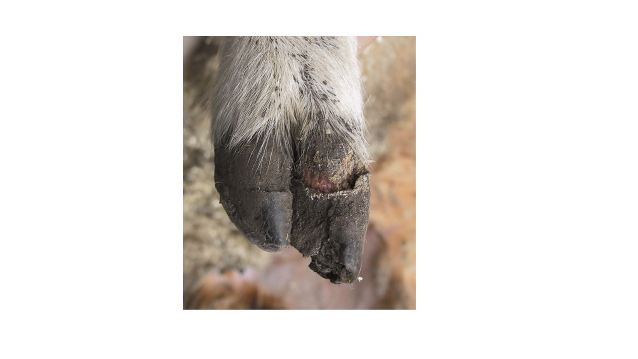
Picture Credit: Wikivet

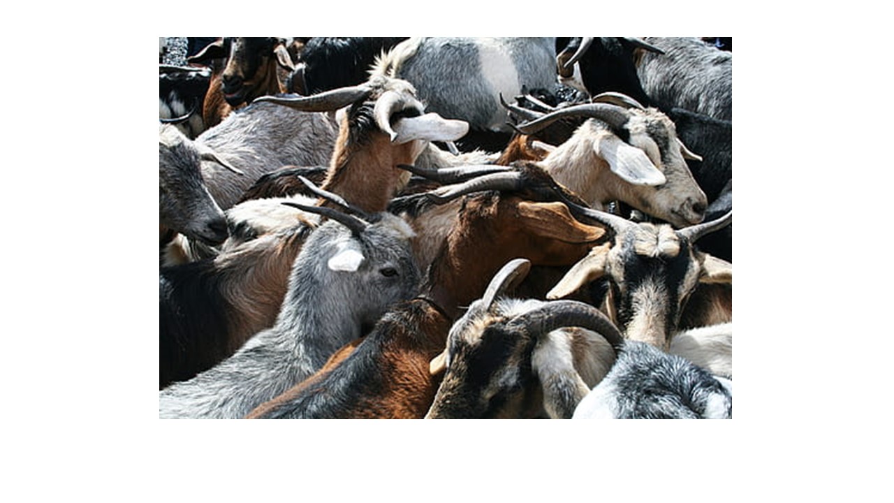
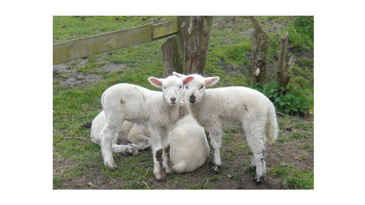
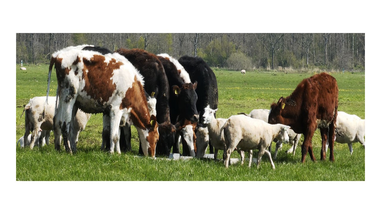
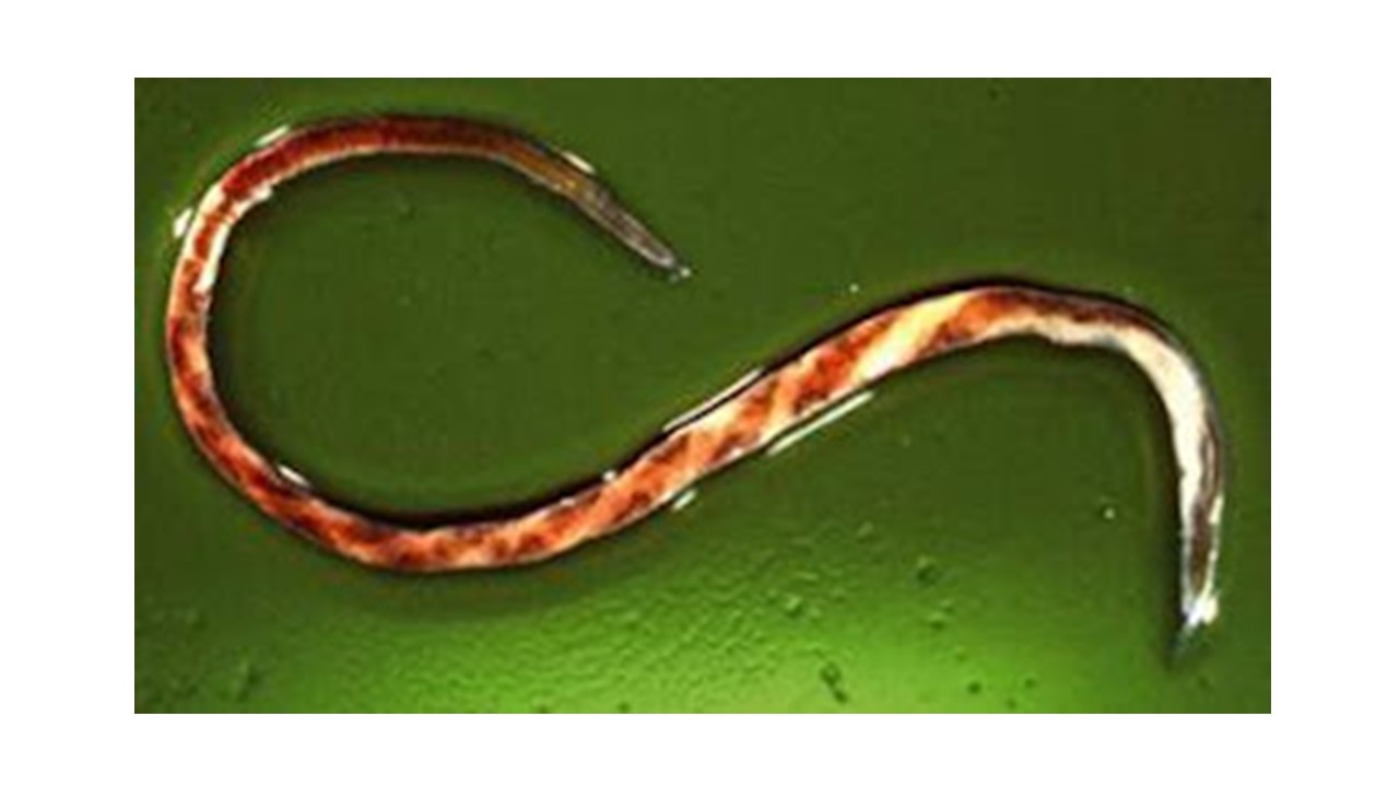
Interdigital dermatitis is a condition that leads to lameness in sheep and goats and is often a predisposing factor to footrot. It is sometimes referred to as Benign Footrot. It is a mixed bacterial infection however, Dichelobacter nodosus has been recognized as the major cause of this condition. Other causal factors of this disease include wet weather, muddy pens, and damp pastures. Diagnosis is based on clinical signs and laboratory testing.
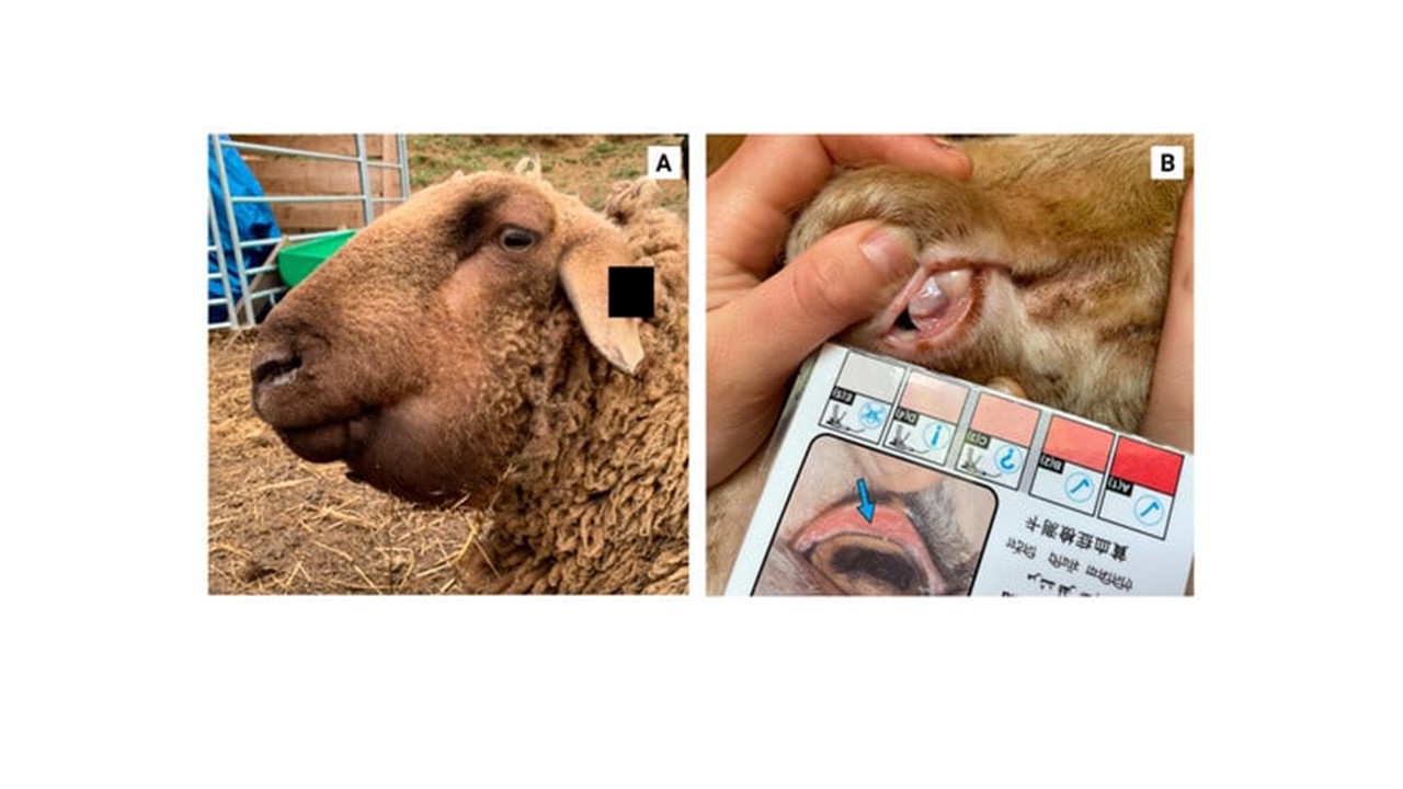
CLINICAL SIGNS

The interdigital skin is red and swollen and may be seen as covered with a thin layer of white exudate. The hoof capsule may be shed in very severe cases. Chronic infections can lead to lameness and formation of misshapen hooves.

TREATMENT, PREVENTION AND CONTROL

The use of topical aerosol antibiotic sprays have proven to be very effective. In farm herds where large numbers have been affected, the whole flock can be treated with 10% Zinc Sulphate solution or 3% formalin in a footbath. High concentrations of formalin greater than 5% can cause irritations. Sheep must be made to stand in a dry area for the formalin or the zinc sulphate to dry on the feet. Footbaths  must be repeated (after a week or two). There is no need to attempt foot trimming.

HAEMONCHOSIS IN RUMINANTS

HAEMONCHOSIS IN RUMINANTS 1280 720 Kingsley Emmanuel Bentum

Photo Credit: Adduci, Isabella, et al. “Haemonchosis in Sheep and Goats, Control Strategies and Development of Vaccines against Haemonchus contortus.” Animals 12.18 (2022): 2339.

Amid the several parasitic infections that plague ruminants, Haemonchus spp has been recognized as one of the many parasites that cause severe damages to the digestive system. This blood-sucking worm is responsible for a lot of losses in cattle, sheep and goat production.

A parasite generally is any organism, that lives in or on another (host) and harms or offers no advantage to the other organism (host).

This worm also commonly referred to as Barber’s Pole worm resides in the abomasum of ruminants (fourth or last chamber of the ruminant’s stomach).  Haemonchus spp has piercing mouthparts that causes extensive damage to the walls of the abomasum. An adult worm is capable of sucking about 0.05mls of blood daily from an affected animal. Hence an animal infected with about 2000 worms will have 100mls of blood loss daily.

There are various species of the blood parasite affecting various ruminant types. Some of the common species of Haemonchus include Haemonchus contortus, Haemonchus similis and Haemonchus placei. There are few records of cross-transmission of the Haemonchus spp between small ruminants and cattle.  Haemonchosis has remained a threat to ruminant production especially in the tropical, subtropical and warm temperate regions of the world, where environmental conditions favour the free-living stages of the parasite.

CLINICAL SIGNS & DIAGNOSIS

In acute conditions, anaemia, agalactia in ewes, submandibular oedema or bottle jaw and death due to blood loss are among the common clinical signs. When the condition becomes chronic, there is progressive weight loss and general weakness. Diagnosis is based on presenting clinical signs, grazing history as well as worm egg count from the laboratory.

TREATMENT & PREVENTION

Consult your veterinarian once you notice similar signs on your farm. Having a good management program to control parasitic infection on your farm is important.

ORF IN RUMIMANTS

ORF IN RUMIMANTS 1280 720 Kingsley Emmanuel Bentum

Orf is a highly transmissible viral disease of sheep and goats with zoonotic potential (i.e. it can be transferred to humans). It has numerous names such as Sore Mouth Disease, Contagious Ecthyma or Scabby Mouth. Orf belongs to the Poxviridae family. Animals get infected through direct contact with affected animals as they interact. Other sources of transmission include from mother to young animals through the teats and through broken skin or wounds as animals graze and feed.  

CLINICAL SIGNS

Lesions normally start as small reddened patches around the mouth, muzzle, teats and legs, which begin to produce a clear fluid. The fluid eventually hardens into a thickened scab which dries up and drops off after 2-3 weeks. The scab in the early stages is firmly attached. Forceful removal of the scab leads to bleeding. When lesions are formed on the udder and teats, nursing mothers may not allow young ones to suckle. Such young ones will require supplemented feed to survive.  

Humans contract the disease when they come in contact with equipment contaminated with the virus. People with close contact to livestock are highly exposed to this virus.

TREATMENT AND PREVENTION

Maintain high biosecurity protocols on farm. Quarantine new animals brought into the farm, as well as affected animals to limit spread of infection. The disease in small ruminants is self-limiting and supportive care is usually given to affected animals. Contact your vet for advice when your animals start exhibiting similar signs.  There are vaccines available, however they are only used in areas where outbreaks have occurred. Vaccine protection is not lifelong but will reduce severity of the disease when it occurs.

DIARRHOEA IN NEWLY BORN RUMINANTS CAUSED BY E.coli

DIARRHOEA IN NEWLY BORN RUMINANTS CAUSED BY E.coli 1280 720 Kingsley Emmanuel Bentum

Diarrhoea in newly born ruminants caused by Escherichia coli (E.coli) remains as one of the common conditions of newborns which sometimes leads to death.

E. coli, is a bacterium which naturally resides in the intestines of living organisms and aid in digestion. There are however some strains of E. coli (Enterotoxigenic E. coli and Enteropathogenic E. coli) that cause severe infection especially in the early lives (normally within the first week) of calves and lambs. The infection is sometimes complicated with other pathogens such as Clostridium perfringens and Rota virus.

Enterotoxigenic E. coli release enterotoxins which disrupts the osmotic balance of the gut cells, leading to excess fluid secretion into the lumen of the gut, resulting in diarrhoea.  

Enteropathogenic E. coli affect the intestinal lining, leading to ulcers and erosions of the intestinal walls. Some of these strains of E. coli release toxins (verotoxin) which enhance the erosions of the intestinal mucosa leading to hemorrhages.

Clinical Signs

Diarrhoea (very clear watery stool or bloody)

– Dehydration

– Death

– Lethargy

– Fever

Treatment and Prevention

– Practice good hygiene especially in delivery pens

– Ensure young animals get colostrum especially during the first week of life. Kids (baby goats)  are however resistant to this infection.

Affected animals are usually given supportive therapy with antibiotics after sensitivity testing has been carried out.

BLACK LEG/ BLACK QUARTER DISEASE

BLACK LEG/ BLACK QUARTER DISEASE 1280 720 Kingsley Emmanuel Bentum

Copyright © 2021 Farm4Trade SRL

This is a highly fatal disease of farm animals including cattle, sheep, goats and horses. It is caused by the bacterium, Clostridium chauvoei (Cl.chauvoei). The disease is characterized by severe swelling and necrosis of affected muscles (especially those of the hind quarters).

Clostridium chauvoei spores can be found in the soil. Drastic changes in the weather such as droughts and floods can expose the bacterial spores which are ingested by animals during feeding. After an animal comes in contact with the bacterium, the organism travels through the digestive system, enters the blood stream and is finally deposited in the muscle and other vital organs such as the spleen and liver. The bacterium can also enter the body through wounds and scratches (especially in small ruminants). Once it enters the body, it produces toxins which are deadly to the host animal.

Signs of Black Leg disease include sudden death of farm animal, fever, depression, lameness (especially the hind quarters) with extensive and painful swelling of the affected limb. Crackling sounds may be heard when affected limbs are touched due to accumulated gas in the muscles (emphysema). Animals usually die within 48 hours after signs of disease are evident.  

Black Leg/Black Quarter disease can be diagnosed through culture, fluorescent antibody test and necropsy.

Treatment of Black Quarter disease is effective when detected early. Contact your veterinarian when you notice similar signs in your farm animals.

To control the spread of this disease, carcasses affected with this disease should be burned completely or buried deeply. Lime or other disinfectants should be sprinkled over the carcass during the burial period.

BRUCELLOSIS (MEDITERRANEAN FEVER/ UNDULANT FEVER) IN SMALL RUMINANTS

BRUCELLOSIS (MEDITERRANEAN FEVER/ UNDULANT FEVER) IN SMALL RUMINANTS 1280 720 Kingsley Emmanuel Bentum

Brucellosis is a highly infectious disease that has the potential of being transferred from animals to humans (zoonotic). It makes it a disease of great public health significance with economic impact.

ETIOLOGY / CAUSATIVE AGENT

It is caused by the bacterium Brucella spp. Brucella has different species including Brucella abortus, Brucella canis, Brucella suis  and Brucella melitensis. These species affect several animals such as cattle, goats, sheep, pigs, dogs, horses and camels. Recent scientific discoveries have identified strains of brucella in red foxes and certain marine animals. Brucella melitensis is the main strain that affects sheep and goats and also has the potential of infecting humans.

MODE of INFECTION

Brucella spp is spread through contact with contaminated birth or uterine fluids, milk and semen from infected animals. Other means of contracting the disease include cuts or scratches through the mucous membranes. 

CLINICAL SIGNS

These may include abortions during the late stages of pregnancy, retained placentas, still births, weak offspring, swollen or inflamed testicles (orchitis) and arthritis. In human, symptoms may include fever, headaches, muscle, and joint pains.

PREVENTION and CONTROL

Brucellosis is difficult to be treated and hence no practical treatment protocol exist in animals. Testing of animals and the elimination of positive reactors is the recommended protocol for control and prevention of the disease. It is important to purchase animals from credible sources and test breeding stock for brucellosis before mating them. After birth discharges should be properly disposed off to reduce spread of infection

BLOAT/ RUMINAL TYMPANY

BLOAT/ RUMINAL TYMPANY 1280 720 Kingsley Emmanuel Bentum

Bloat is an uncomfortable condition in ruminants marked by an over distension of the rumen (the first of the four divisions of stomach in ruminants). There are microbes which are naturally found in the rumen and aid in the fermentation process of feed. Gas naturally produced due to this process is expelled by eructation or burping. Bloat occurs when there is any form of hindrance to the normal release of gas from the rumen.

TYPES OF BLOAT

Free-Gas Bloat: This is a less common type, and it occurs due to the oesophagus being blocked by a foreign object such as a lump of feed. There are other medical conditions such as tetanus, tumors and hypocalcaemia (reduced calcium levels in the blood) which can affect the movement of the oesophagus, leading to this condition.

Frothy Bloat: This is the most common type of bloat. It occurs usually during the onset of the rainy season when there is a lot of fresh highly proteinous forage such as legumes. These plants are rapidly digested leading to the formation of a layer of entrapped gasses in the form of foam, which makes it difficult to be released from the rumen.

CLINICAL SIGNS

  • A highly distended left abdomen
  • Sudden death
  • Difficulty in breathing (dyspnea)
  • Death can occur within 4 hours after signs of bloat begin to show due to an impairment of normal respiration.

DIAGNOSIS

Diagnosis is usually based on history and physical examination. The use of the stomach tube is useful in distinguishing between free-gas and frothy bloat

TREATMENT and PREVENTION

  1. A stomach tube or Trocar and Cannula is used to release excess gas from the rumen. In life threatening cases a surgical procedure may be needed.
  2. Antifoaming products such as Poloxalene, vegetable and mineral oils have proven to be effective.

Avoid grazing animals on high-risk pastures. Diet of animals fed in stalls should contain a balance of grains and roughage. You may consider providing your farm animals antifoaming agents especially during the rainy season when there is a lot of lush vegetation. Ensure that feed or pastures are always free from foreign materials such as plastics and stones.

HAEMONCHOSIS

HAEMONCHOSIS 1280 720 Kingsley Emmanuel Bentum

Photo Credit: http://www.sheep101.info/201/parasite.html

Amid the several parasitic infections that plague ruminants, Haemonchus spp has been recognized as one of the many parasites that cause severe damages to the digestive system. This blood-sucking worm is responsible for a lot of losses in cattle, sheep and goat production.

A parasite generally is any organism, that lives in or on another (host) and harms or offers no advantage to the other organism (host).

This worm also commonly referred to as Barber’s Pole worm resides in the abomasum of ruminants (fourth or last chamber of the ruminant’s stomach).  Haemonchus spp has piercing mouthparts that causes extensive damage to the walls of the abomasum. An adult worm is capable of sucking about 0.05mls of blood daily from an affected animal. Hence an animal infected with about 2000 worms will have 100mls of blood loss daily.

There are various species of the blood parasite affecting various ruminant types. Some of the common species of Haemonchus include Haemonchus contortus, Haemonchus similis and Haemonchus placei. There are few records of cross-transmission of the Haemonchus spp between small ruminants and cattle.  Haemonchosis has remained a threat to ruminant production especially in the tropical, subtropical and warm temperate regions of the world, where environmental conditions favour the free-living stages of the parasite.

CLINICAL SIGNS & DIAGNOSIS

In acute conditions, anaemia, agalactia in ewes, submandibular oedema or bottle jaw and death due to blood loss are among the common clinical signs. When the condition becomes chronic, there is progressive weight loss and general weakness. Diagnosis is based on presenting clinical signs, grazing history as well as worm egg count from the laboratory.

TREATMENT & PREVENTION

Consult your veterinarian once you notice similar signs on your farm. Having a good management program to control parasitic infection on your farm is important.